Software-based CatLet score algorithm
The CatLet score calculation is feasible by use of a computer program consisting of sequential and interactive self-guided questions (available at http://www.catletscore.com).
The algorithm includes 15 main questions. The first question includes 3 sub questions: RCA dominance, Dx size and LAD length. Answering these 3 sub-questions will define one of 54 types of coronary artery circulations proposed in this angiographic scoring system (different from the SYNTAX score) (13). The second question includes two options: option 1 is for recording normal coronary artery anatomy; and option 2 (most important) is for evaluating the lesions. Question 3 addresses lesion-based unlimited segment selection. Question 4 addresses 2 native or in-stent lesion types, including non-occlusions and acute total occlusion (ATO).
For non-occlusion or ATO lesions, questions 5-12 address the lesion-based evaluation, including involved segments, calcifications, angulations, and thrombus, which are most similar to those adopted in the SYNTAX score (13), but differ in only qualitatively recording these adverse characteristics as “Yes” or “No” in our scoring system (table 7). Question 12 addresses scoring modification if necessary. Upon completion of question 12, the algorithm proceeds to the last question or to another cycle of “add another lesion”. The last question of the CatLet score algorithm, diffuse disease/small vessels, similar to those in the SYNTAX score, is also only qualitatively recorded (table 7). For a patient with coronary artery disease, it takes around 5-10 minutes to process a patients depending on the complexity and severity of coronary artery disease.
Table 7. Scoring and describing of lesion’s adverse characteristics.
Diameter reduction | |
-Total occlusion | ×5 |
-Significant lesion (50-99% diameter reduction) | ×2 |
Total occlusion | |
-Native non-occlusion | Yes or No |
-In-stent non-occlusion | Yes or No |
-Native acute total occlusion (ATO) | Yes or No |
-In-stent ATO | Yes or No |
"Culprit" lesion judgment | Yes or No |
Trifurcations | Yes or No |
Bifurcations | Yes or No |
Medina types | Yes or No |
Angulation<70 | Yes or No |
Aorto ostial stenosis | Yes or No |
Severe tortuosity | Yes or No |
Length > 20 mm | Yes or No |
Heavy calcification | Yes or No |
Thrombus | Yes or No |
Anatomic description of native coronary system | Yes or No |
In the CatLet score, only the lesion's stenosis was scored, and the lesion's remaining adverse characteristics were instead qualitatively recorded. CTO is defined an occluded coronary segment with TIMI flow 0 for ≥3 months duration. Definitions for lesions' adverse characteristics are the same as listed in the Syntax score (13). In-stent lesions are defined as angiographic diameter stenosis >50% at the stent segment or its adjacent edges within 5 mm (15).
Scoring of a sample patient according to the CatLet angiographic scoring system
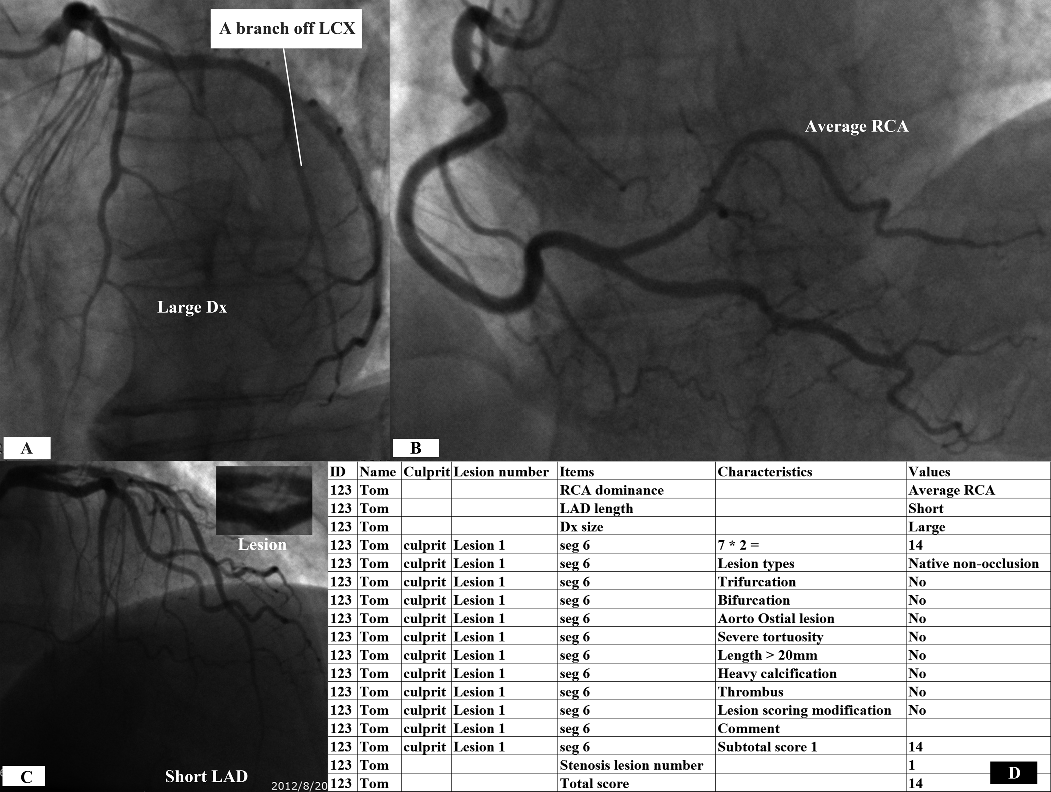
As shown in Figure 16, coronary circulation pattern was first evaluated overall according to LAD length, Dx size and RCA size (Figure 16A-C) before the non-occlusive lesion on proximal LAD (segment 6) was scored. Average RCA was judged according to the presence of a significant branch arising from LCX. The lesion was amplified right upper in the Figure 16C. Adverse non-stenotic characteristics pertinent to the lesion were no longer scored and qualitatively recorded as Yes or No. Detailed information on the lesion was displayed in Figure 16D.

Figure 16. Scoring of a sample patient according to the CatLet score algorithm.
References:
1. Cannon PJ, Dell RB, Dwyer EM, Jr. Measurement of regional myocardial perfusion in man with 133 xenon and a scintillation camera. J Clin Invest 1972;51:964-77.
2. Dwyer EM, Jr., Dell RB, Cannon PJ. Regional myocardial blood flow in patients with residual anterior and inferior transmural infarction. Circulation 1973;48:924-35.
3. Ganz W, Tamura K, Marcus HS, Donoso R, Yoshida S, Swan HJ. Measurement of coronary sinus blood flow by continuous thermodilution in man. Circulation 1971;44:181-95.
4. Holman BL, Adams DF, Jewitt D, et al. Measuring regional myocardial blood flow with 133Xe and the Anger camera. Radiology 1974;112:99-107.
5. Klocke FJ. Coronary blood flow in man. Prog Cardiovasc Dis 1976;19:117-66.
6. Klocke FJ, Bunnell IL, Greene DG, Wittenberg SM, Visco JP. Average coronary blood flow per unit weight of left ventricle in patients with and without coronary artery disease. Circulation 1974;50:547-59.
7. Mymin D, Sharma GP. Total and effective coronary blood flow in coronary and noncoronary heart disease. J Clin Invest 1974;53:363-73.
8. Ross RS, Ueda K, Lichtlen PR, Rees JR. Measurement of Myocardial Blood Flow in Animals and Man by Selective Injection of Radioactive Inert Gas into the Coronary Arteries. Circ Res 1964;15:28-41.
9. Gallik DM, Obermueller SD, Swarna US, Guidry GW, Mahmarian JJ, Verani MS. Simultaneous assessment of myocardial perfusion and left ventricular function during transient coronary occlusion. J Am Coll Cardiol 1995;25:1529-38.
10. Cerqueira MD, Weissman NJ, Dilsizian V, et al. Standardized myocardial segmentation and nomenclature for tomographic imaging of the heart. A statement for healthcare professionals from the Cardiac Imaging Committee of the Council on Clinical Cardiology of the American Heart Association. Circulation 2002;105:539-42.
11. Austen WG, Edwards JE, Frye RL, et al. A reporting system on patients evaluated for coronary artery disease. Report of the Ad Hoc Committee for Grading of Coronary Artery Disease, Council on Cardiovascular Surgery, American Heart Association. Circulation 1975;51:5-40.
12. The ARTS (Arterial Revascularization Therapies Study): Background, goals and methods. Int J Cardiovasc Intervent 1999;2:41-50.
13. Sianos G, Morel MA, Kappetein AP, et al. The SYNTAX Score: an angiographic tool grading the complexity of coronary artery disease. EuroIntervention 2005;1:219-27.
14. Ormiston JA, Kassab G, Finet G, et al. Bench testing and coronary artery bifurcations: a consensus document from the European Bifurcation Club. EuroIntervention 2018;13:e1794-e803.
15. Kuntz RE, Baim DS. Defining coronary restenosis. Newer clinical and angiographic paradigms. Circulation 1993;88:1310-23.
发表评论:
◎Welcome to participate in the discussion, please post your views here and exchange your views.